
The human eye is a marvel of biological engineering that allows us to perceive the world around us. This guide will take you through the various components of the human eye, describe their functions, and illustrate how these parts work in harmony to create vision. Detailed diagrams and in-depth explanations accompany each section to enhance your understanding.
Structure of the Human Eye
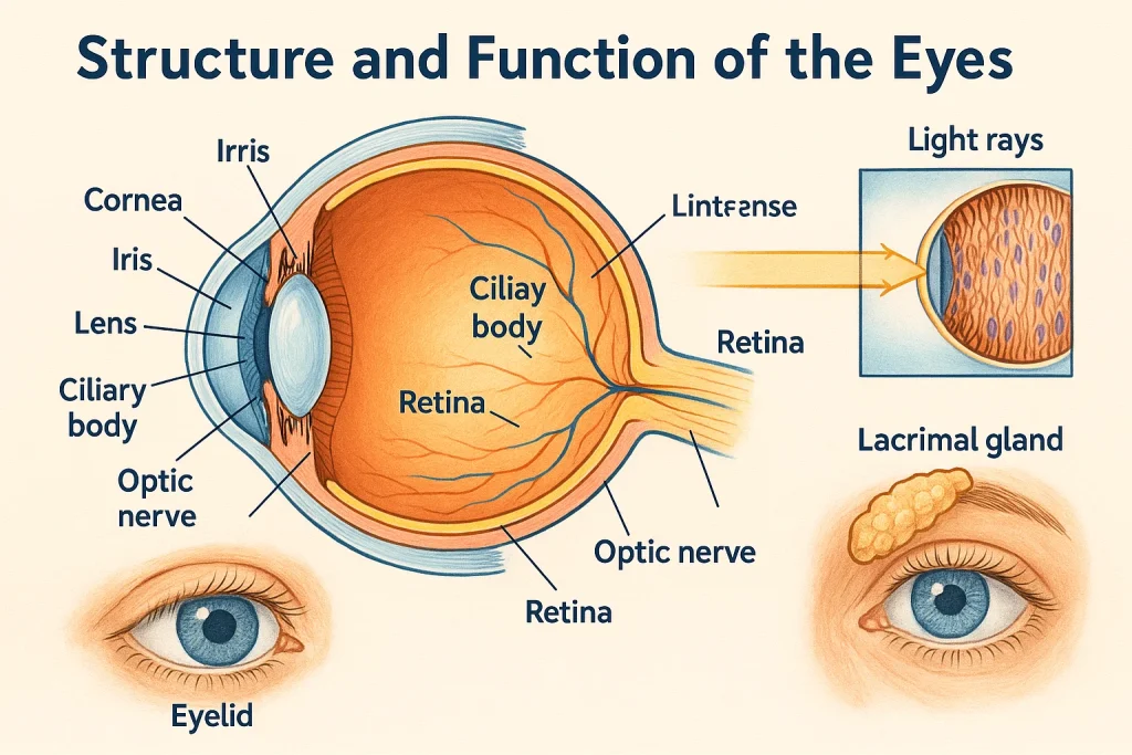
The human eye is roughly spherical and comprises several distinct parts, each playing a crucial role in vision. Below, we break down these components, explain their functions, and provide corresponding diagrams for clearer visualization.
1. Cornea
- Description:
The cornea is the transparent, dome-shaped outer layer that covers the front of the eye. It is the eye’s first refractive medium, directing and bending incoming light. - Function:
It contributes approximately 65-75% of the eye’s optical power by refracting light to begin focusing it on the retina.
2. Iris
- Description:
The iris is the colored part of the eye—a muscular structure situated behind the cornea. - Function:
It regulates the size of the pupil, controlling the amount of light that enters the eye. In bright light, the iris constricts the pupil; in low light, it dilates.
3. Pupil
- Description:
The pupil is the central opening in the iris. - Function:
It acts similarly to the aperture of a camera, controlling the light intensity that reaches the inner eye.
4. Lens
- Description:
Positioned immediately behind the pupil, the lens is a flexible, biconvex structure. - Function:
It fine-tunes the focus of light onto the retina through a process called accommodation—changing shape to adjust focus for near or far objects.
5. Retina
- Description:
The retina is a thin layer of tissue lining the back of the eye. - Function:
It contains photoreceptor cells—rods for low-light vision and cones for color and sharp vision—that convert light into electrical signals.
6. Optic Nerve
- Description:
The optic nerve is a bundle of nerve fibers that transmits visual information from the retina to the brain. - Function:
It acts as the communication link, sending processed visual messages to the brain where they are interpreted as images.
7. Ciliary Muscles
- Description:
These are ring-shaped muscles that encircle the lens. - Function:
They control the shape of the lens by contracting or relaxing, a process crucial for adjusting focus on objects at various distances (accommodation).
8. Sclera
- Description:
The sclera is the opaque, white outer layer of the eyeball. - Function:
It protects the inner components of the eye and provides structural support.
9. Aqueous and Vitreous Humor
- Description:
- Aqueous humor: A clear fluid between the cornea and lens.
- Vitreous humor: A gel-like substance that fills the main cavity of the eye.
- Function:
These fluids help maintain the shape of the eye, provide nutrients, and assist in light refraction.
Function of the Human Eye
Understanding how the eye works involves exploring the sequence of events from light entry to image formation and processing by the brain.
How Vision Occurs: A Step-by-Step Process
- Light Entry and Refraction through the Cornea:
- Process: Light first passes through the transparent cornea, which refracts and directs it toward the lens.
2. Pupil Adjustment by the Iris:
- Process: The iris adjusts the pupil size to control the intensity of light reaching the inner eye.
3. Focusing by the Lens:
- Process: The lens fine-tunes the focus through accommodation, ensuring that light rays converge accurately on the retina.
4. Image Formation on the Retina:
- Process: As light is focused on the retina, photoreceptor cells convert the light into electrical impulses.
5.Transmission via the Optic Nerve:
- Process: The generated electrical impulses are transmitted to the brain through the optic nerve.
Common Vision Defects and Their Corrections
Understanding the normal function of the eye also helps in identifying when things go awry. Here are some common vision defects:
Myopia (Nearsightedness)
- Cause: Light is focused in front of the retina.
- Correction: Concave lenses help diverge light rays.
Hypermetropia (Farsightedness)
- Cause: Light focuses behind the retina.
- Correction: Convex lenses help converge light rays.
Astigmatism
- Cause: Irregular curvature of the cornea or lens leads to distorted images.
- Correction: Cylindrical lenses are used to correct the vision.
Presbyopia
- Cause: Age-related loss of lens flexibility.
- Correction: Use of reading glasses or bifocal lenses.
Conclusion
The human eye is a complex yet beautifully coordinated organ that plays an essential role in how we experience the world. Each component—from the cornea to the retina—has a distinct structure and function, working collectively to convert light into meaningful visual information. With the aid of detailed diagrams and step-by-step explanations, this guide aims to simplify the subject, making it accessible and engaging for students.
FAQs
Q1: What is the primary function of the retina?
A1: The retina converts incoming light into electrical signals using photoreceptor cells (rods and cones), which are then sent to the brain via the optic nerve.
Q2: How does the iris regulate the amount of light entering the eye?
A2: The iris adjusts the pupil size, controlling the volume of light that passes through to the lens and retina.
Q3: What role does the lens play in vision?
A3: The lens fine-tunes the focus of light by changing its shape (accommodation) to ensure that a clear image is formed on the retina.
Q4: How do corrective lenses help with vision defects like myopia and hypermetropia?
A4: Concave lenses diverge light, correcting myopia by moving the focal point back onto the retina, while convex lenses converge light, correcting hypermetropia by moving the focal point forward.